
Diabetic Blisters can be startling to find, especially if they appear overnight. These fluid-filled sacs often look like burns but usually form without a clear injury. Most are painless and heal with careful protection. This guide explains causes, signs, and safe care, with a focus on prevention and when to seek help.
Key Takeaways
- Typical features: sudden, painless, sterile blisters on feet, legs, or hands.
- Core priorities: protect skin, reduce pressure, and watch for infection.
- See a clinician promptly if redness, warmth, pain, or fever develop.
- Prevention relies on foot checks, good footwear, and glucose control.
Understanding Diabetic Blisters
Clinicians often refer to these as bullosis diabeticorum, a rare blistering condition seen mostly in long-standing diabetes. Lesions tend to appear on weight-bearing or trauma-prone sites. The blisters are tense, filled with clear fluid, and usually non-tender. They may occur singly or in clusters and often resolve over weeks if protected from friction.
The exact mechanism is not fully understood. Proposed drivers include microvascular changes, neuropathy-related unnoticed trauma, and glycation effects on skin integrity. Because the fluid is typically sterile, routine antibiotics are not needed unless infection is suspected. For a concise clinical overview, see this peer-reviewed summary from StatPearls (clinical overview).
Causes and Risk Factors
Researchers group diabetic blisters causes into three broad factors: mechanical stress, impaired healing, and microvascular compromise. Minor, unrecognized friction from shoes, bed linens, or braces can trigger localized skin separation. Poor glycemic control may weaken skin structure and slow recovery. Peripheral neuropathy reduces pain signaling, so injuries go unnoticed and unprotected.
Other contributors include dehydration, ill-fitting footwear, and previous foot deformities like hammertoes or Charcot changes. Autonomic neuropathy may dry the skin, making it fragile and easy to shear. When blisters recur, it can signal broader metabolic imbalance. For context on recognizing overall imbalance patterns, see Signs Of Uncontrolled Diabetes for pattern recognition and Diabetic Neuropathy for nerve-related risk factors.
Symptoms, Diagnosis, and Differentials
Typical findings include sudden-onset, tense blisters with clear fluid on the feet, lower legs, or hands. Many people report no pain, fever, or preceding trauma. Clinicians look for intact surrounding skin, minimal inflammation, and lack of pus. Because infection changes management, providers assess warmth, erythema, and tenderness closely. Bullosis diabeticorum symptoms overlap with other blistering diseases, so careful examination matters.
Differentials include friction blisters, burns, insect bites, herpetic lesions, and autoimmune bullous disorders. Bullous pemphigoid often shows widespread itch and urticarial plaques, while epidermolysis bullosa acquisita presents with scarring. Cultures are considered if infection is likely. When the presentation is atypical, dermatology may recommend a small biopsy or direct immunofluorescence to rule out autoimmune causes. For broader context on skin disease patterns in diabetes, see Diabetes Skin Problems for key differentiators and Diabetes And Fungal Infections for common infectious look-alikes.
What Clinicians Look For
Examination usually documents number, size, and location of blisters, plus skin perfusion, sensation, and footwear fit. Imaging is rarely needed unless deep infection is suspected. Providers may photograph lesions to monitor changes in size, color, and surrounding skin. They also check pulses, monofilament sensation, and pressure points. This baseline helps guide offloading, dressing choices, and follow-up intervals. Photographs can also support education and earlier detection during telemedicine visits.
Where They Occur and What Pictures Show
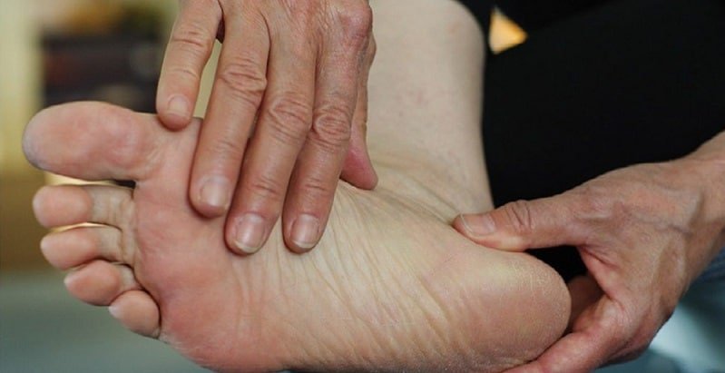
Most lesions appear on the feet and lower legs, with fewer on the hands and forearms. Visual references help people recognize typical morphology, distribution, and surrounding skin. When people search for pictures of diabetic blisters, they usually want to confirm whether a new lesion is consistent with this condition. Use images to guide self-checks, but avoid self-diagnosis if signs of infection appear.
Early foot images often show intact roofs with clear fluid, little redness, and minimal scaling. By contrast, ulcer photos show open skin loss, drainage, or necrotic tissue. If a lesion looks more like an ulcer, especially on weight-bearing areas, seek care. For a focused comparison of open lesions, see Diabetic Foot Ulcers for distinguishing features and complications.
Home Care, Dressings, and Monitoring
Conservative measures form the backbone of diabetic blisters treatment. Keep the blister roof intact if possible; it protects against bacteria and reduces pain. Cover with a nonadherent dressing and change it if damp or dirty. Reduce friction with soft padding and avoid tight footwear or repetitive pressure. Inspect the area daily for redness, clouded fluid, or increased tenderness.
Clean surrounding skin with mild soap and water. Do not puncture at home unless a clinician instructs you. Offload pressure using cushioned insoles, felt padding, or temporary activity changes. If the roof tears, trim only loose, nonviable skin under clinical guidance and keep the base moist, not wet. For background on delayed repair biology, see Wound Healing Process to understand why careful moisture balance matters. Note: Apply topical antibiotics only if directed by a clinician evaluating signs of local infection.
When to Seek Urgent Care and Complications
People often ask, are diabetic blisters dangerous. Most are not, but they can allow bacteria to enter if the roof breaks. Seek urgent care for spreading redness, warmth, foul odor, purulent drainage, increasing pain, or fever. Red streaks up the leg, rapidly enlarging wounds, or inability to bear weight also warrant prompt evaluation. These findings suggest evolving cellulitis or deeper infection that needs medical treatment.
Foot infections progress quickly in diabetes due to impaired immunity and circulation. Early evaluation reduces risks of tissue loss and hospitalization. Routine foot self-checks catch issues earlier. For practical steps on skin and foot checks, the CDC provides concise foot care guidance to structure a safe home routine.
Professional Options and Prevention
In clinic, bullosis diabeticorum treatment is tailored to blister integrity and infection risk. Intact blisters are usually protected, not deroofed. If the blister is very tense and painful, a clinician may aspirate with sterile technique while preserving the roof. Open lesions are cleaned, conservatively debrided if needed, and covered with a nonadherent dressing. Offloading devices or felted foam reduce pressure. Antibiotics are reserved for confirmed or strongly suspected infection.
Prevention focuses on three pillars: skin protection, pressure reduction, and metabolic stability. Wear properly fitted shoes, moisture-wicking socks, and consider orthotics for deformities. Hydrate and apply fragrance-free emollients to reduce shear. Daily checks help catch small changes before they escalate. For medication background that supports glucose targets, see Common Diabetes Medications for therapy overviews. For broader skin complication context, review the American Diabetes Association’s summary on skin complications and integrate its prevention points into daily routines.
Foot-Specific Care and Footwear Strategies
Because most lesions occur on weight-bearing areas, targeted support helps. Diabetic foot blister treatment emphasizes pressure redistribution with cushioned insoles, metatarsal pads, or temporary offloading boots if needed. Consider a gradual break-in schedule for new shoes, checking skin after each wear. Socks should be seamless, moisture-wicking, and appropriately padded to limit shear forces.
Schedule regular foot checks with your care team, especially if you have neuropathy or previous ulcers. A podiatrist can advise on callus control, nail care, and protective footwear. For guidance on team roles and referrals, see What Is a Podiatrist for scope of practice details and Foot Screening For Diabetes for structured exam intervals and risk stratification.
Hands and Legs: Special Situations
Blistering on the hands often reflects unrecognized friction from tools, exercise gear, or mobility aids. Protective gloves, padded grips, and frequent skin checks reduce risk. Over the shins and calves, rubbing from braces or socks can shear fragile skin. Adjust fit, add soft interfaces, and elevate legs if swelling increases tension. Monitor carefully if you have sensory loss.
Any blister that becomes warm, painful, or cloudy requires prompt assessment. If limbs are swollen or discolored, clinicians may evaluate peripheral arterial disease or venous insufficiency. These conditions complicate healing and may require vascular input. For an integrated skin-health perspective and practical routines, review Diabetes Skin Problems: What You Need To Know to deepen prevention strategies at home.
Recap
Sudden, painless blistering in diabetes is usually sterile and self-limited when protected. Keep the roof intact, reduce friction, and watch closely for infection. Seek timely care for redness, warmth, drainage, or fever. Consistent foot checks, footwear optimization, and glucose stability help prevent recurrences. For day-to-day routines, align home practices with trusted clinical resources and follow your care team’s advice.
Tip: Photograph new lesions with date stamps. Images help you and your clinician track change, spot early infection, and adjust offloading.
This content is for informational purposes only and is not a substitute for professional medical advice.
