
Key Takeaways
Diabetes and Wound Healing involves delayed repair due to high glucose, impaired blood flow, and nerve damage.
- Monitor blood glucose closely; stable control supports tissue repair.
- Catch small injuries early; act before ulcers deepen.
- Offload pressure and keep wounds clean and moist.
- Check feet daily; address calluses, blisters, and fungal issues.
Diabetes and Wound Healing
High blood sugar disrupts normal repair by slowing inflammation resolution and collagen rebuilding. Nerve damage (neuropathy) reduces pain sensation, so minor trauma goes unnoticed. Poor circulation from small-vessel disease (microangiopathy) limits oxygen delivery to tissues. Together, these factors increase infection risk and prolong closure times.
This section explains key mechanisms in clinical terms and plain language. For a broader overview of the wound healing process in diabetes, see Affect Wound Healing Process for foundational context and definitions.
Pathophysiology: What Changes at the Tissue Level
Hyperglycemia increases advanced glycation end-products (glycation), which stiffen proteins and impair cell signaling. White blood cells function less effectively, reducing bacterial killing. Capillaries thicken, dropping perfusion. These changes weaken granulation tissue and slow epithelial closure.
Clinicians often ask how does diabetes affect wound healing pathophysiology because the drivers determine care. Neuropathy (nerve damage) promotes repetitive trauma, while ischemia (poor blood flow) starves tissues of oxygen. Biofilm formation on chronic wounds further impedes closure. For deeper mechanisms and summaries, the NIH review on wound healing provides balanced background and diagrams.
Common Lesions and Staging
Diabetes increases risk for foot ulcers, pressure injuries, cellulitis, and surgical site problems. Toe deformities and calluses concentrate pressure, creating skin breakdown. Venous and arterial disease may coexist, complicating diagnosis. Document size, depth, and tissue type at each visit to track change.
Clinicians categorize lesions by depth, infection, and ischemia. Discuss types of diabetic wounds with your care team to target treatment. For staging basics and red flags, see Guide to Diabetic Foot Ulcers for a structured walk‑through of severity scales.
Early Recognition and What Pictures Mean
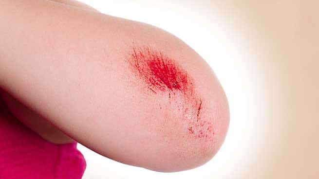
Early photographs often show subtle redness, shiny skin, or a small callused crater. Shallow ulcers may appear clean yet hide deeper tissue risk. Color changes, undermining edges, and drainage patterns suggest infection or ischemia. Use consistent lighting and distance when tracking changes over time.
Searches for diabetic foot ulcer early stage pictures aim to understand when to act. Visual cues, however, cannot replace hands‑on assessment of warmth, tenderness, and pulses. Because delayed care raises amputation risk, review limb‑threatening signs in Why Diabetics Lose Limbs for context on progression and prevention.
Home Care and Clinical Management
First steps focus on cleansing with saline, removing loose debris, and maintaining a moist environment with appropriate dressings. Offload pressure using footwear, felt pads, or casts as advised. Do not use caustic agents like full‑strength iodine on granulating tissue. Elevate when swollen, and protect surrounding skin with barrier creams.
Know how to heal diabetic wounds with a team approach. Monitor glucose consistently; for fingerstick supplies guidance, see Accu Chek Lancets FastClix to understand lancet options for routine checks. Treat infections promptly and avoid self‑lancing abscesses. For localized cellulitis background and complications, read Cellulitis and Diabetes to align symptoms with timing of care. When antibiotics are considered, practice parameters from the IDSA diabetic foot infection guidelines explain culture timing and agent selection.
Speeding Recovery: Practical Tips
Address modifiable barriers systematically. Tight glucose control reduces infection risk and supports collagen formation. Optimize protein intake, hydration, and micronutrients within your meal plan. Evaluate medications (like steroids) that may slow closure, and adjust only under clinical supervision.
People often ask how can a diabetic wound heal faster without risky shortcuts. Offloading, smoking cessation, and edema control usually bring the greatest gains. For nutrition‑adjacent context on antioxidants, see Vitamin C and Diabetes for balanced evidence on supplementation considerations. Some advanced options include negative pressure therapy, cellular dressings, or hyperbaric oxygen, typically reserved for selected cases.
After Procedures: Surgical Incisions
Glycemic variability increases infection and dehiscence risk following operations. Coordinate pre‑ and post‑op plans with your surgeon and diabetes clinician. Inspect incision edges daily for separation, warmth, or unexpected drainage. Keep the site dry until cleared to shower or change dressings.
Concerns about diabetes wound healing after surgery are common and valid. Discuss timing for resuming activity and footwear to reduce stress on sutured areas. Ask when to remove steri‑strips, and what symptoms require urgent review. If fever, spreading redness, or severe pain occurs, seek prompt assessment.
Medications, Dressings, and Ointments
Topicals support moisture balance and bacterial control. Choose non‑adherent dressings that maintain a humid environment without maceration. Silver or iodine‑impregnated products may help bioburden in specific cases. Enzymatic or autolytic debridement can reduce non‑viable tissue when sharp debridement is unsuitable.
Systemic antibiotics target clinically infected wounds; culture guidance steers selection. For product information and indications, review Cephalexin as a commonly used oral option under clinician advice. Foot fungus can worsen skin breaks; for context on antifungals, see Terbinafine or Fluconazole when providers suspect tinea or yeast involvement. Always confirm compatibility with your current medicines.
How Long Healing Takes
Timeframes vary with depth, blood flow, infection, and offloading. Small superficial wounds may close within weeks under optimal conditions. Deeper ulcers, ischemic limbs, or persistent pressure often extend recovery. Thorough assessment and consistent care practices are the best predictors of progress.
Patients ask how long does a diabetic wound take to heal because planning matters. Track weekly changes in size and tissue quality. If measurements stagnate over four weeks, reevaluate offloading, perfusion, and infection status. For vascular contributors and diagnostic steps, explore Peripheral Artery Disease and Diabetes to understand testing and referral timing.
Prevention and Daily Foot Care
Inspect both feet daily, including between toes and under the heel. Use a mirror or assistance if needed. Address corns and calluses professionally rather than self‑paring. Keep skin moisturized, but avoid creams between toes to limit excess moisture.
Trim nails straight across, and choose socks without tight bands. Break in new shoes gradually to prevent hot spots. Replace worn insoles that concentrate pressure. For broader reading across related topics, browse Diabetes Articles to connect foot care with overall metabolic health.
When to Seek Help
Seek care urgently for spreading redness, fever, foul odor, deep probing, or sudden numbness. New black discoloration, rest pain, or cold toes suggest ischemia. Significant swelling, purulent drainage, or severe pain also warrants rapid review. Do not delay if symptoms escalate.
Preventive visits reduce complications. Annual comprehensive foot exams detect neuropathy and vascular disease early. For routine hygiene and skin topics, see Dermatology Articles for supportive context on rashes and fungal care. The ADA foot care guidance outlines periodic screening and footwear recommendations.
Recap
Diabetes alters wound biology through glucose effects, impaired perfusion, and neuropathy. Early detection, offloading, and infection control matter most. Pair consistent glucose management with structured wound checks and timely reviews.
Tip: Keep a simple wound log with photos, size, and dressing type. Small trends often reveal the next best step.
This content is for informational purposes only and is not a substitute for professional medical advice.
