
Key Takeaways
- High glucose slows repair and increases infection risk.
- Nerve damage and poor blood flow delay tissue growth.
- Daily foot checks and offloading prevent new ulcers.
- Early care and cultures guide better treatment choices.
Understanding how wound healing and diabetes intersect helps you spot issues earlier and act promptly. This guide explains why wounds can stall, what signs to track, and which day-to-day steps may reduce complications over time.
Wound Healing and Diabetes: What Changes Biologically
Diabetes alters each stage of normal repair, from inflammation to remodeling. Hyperglycemia (high blood sugar) impairs white blood cell chemotaxis, weakens bacterial killing, and fuels biofilm (slimy bacterial layer) formation. Microvascular disease limits oxygen delivery, while ischemia (low blood flow) slows collagen deposition and angiogenesis (new blood vessel growth).
Peripheral neuropathy (nerve damage) reduces protective sensation, so repetitive pressure goes unnoticed. That pressure produces deeper tissue injury and callus, especially in the midfoot. At a systems level, glycation end-products stiffen tissues and reduce elasticity, making reopening more likely. For deeper mechanisms and practical implications, see Diabetes and Wound Healing for a stepwise overview.
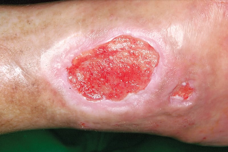
Common Diabetic Wound Patterns on Feet, Legs, and Hands
Feet bear most ulcers due to pressure points, tight footwear, and reduced sensation. Plantar forefoot, under the first and fifth metatarsal heads, and the heel are frequent sites. Lower-leg wounds often follow edema, venous disease, or minor trauma from scratching dry skin. Hands are less common, but burns and cuts may linger, especially around fingertips.
Clinicians often classify the types of diabetic foot ulcer by depth, infection, and ischemia. Neuropathic ulcers occur on pressure points with callus, while neuroischemic ulcers appear on the edges with fragile skin. If scaly plaques or ring-shaped rashes complicate healing, fungal coinfections may be present; for persistent cases, topical options like Ketoconazole are sometimes used for fungal skin involvement when prescribed.
Tip: Address swelling early. Compression for venous disease may help leg wounds when arterial flow is adequate, guided by a clinician.
Timelines, Staging, and Monitoring Progress
Healing rates vary with glucose control, blood flow, depth, and bacterial burden. In practical terms, ask: how long does a diabetic wound take to heal? Superficial abrasions may close within weeks under optimal care, while deep infected ulcers can persist for months. Tracking length, width, depth, and undermining weekly helps determine real progress and adjust care.
Photographs and consistent measurement points allow objective comparisons over time. If a wound fails to reduce area by about 40%–50% within four weeks of appropriate care, reassess offloading, debridement (removal of dead tissue), and infection control. For a plain-language overview of diabetic foot ulcers, MedlinePlus provides helpful context on staging and management.
Early Warning Signs You Can See and Feel
Subtle changes often appear before full ulceration. Look for hot spots, callus buildup, bleeding into callus, shiny swollen skin, or a sudden change in shoe comfort. Early-stage redness with tenderness, especially near bony prominences, warrants pressure relief and inspection for hidden cracks or blisters.
Leg wounds may start as small crusted areas that fail to close, eventually forming diabetic sores on legs treatment plans focused on offloading, edema control, and infection checks. If new neuropathic symptoms emerge, review glucose patterns and consider evaluation of sensation and vibration. For broader skin concerns across conditions, our Dermatology articles explain why rashes and dryness complicate barrier function.
Daily Care, Dressings, and Prevention Basics
Daily habits drive outcomes. Clean with saline or mild soap, keep skin moisturized around but not inside the wound, and use dressings that maintain balanced moisture. Offloading with a boot, felt padding, or custom insole reduces repetitive trauma. Ask a clinician about debridement frequency to control slough and biofilm.
If you wonder how to heal diabetic wounds, start with consistent offloading, glucose management, and early infection detection. Good glycemic control (blood sugar management) supports leukocyte function and collagen synthesis; for therapy background, see our Premixed Insulin Guide when discussing insulin options with your care team. Routine foot care reduces risk; the CDC’s foot care tips summarize simple, protective steps.
Foot checks should include the soles, between toes, and shoes’ interiors. Replace tight or worn footwear and consider moisture-wicking socks. For broader lifestyle context across metabolic health, see Symptoms, Causes, and Prevention for foundational strategies that support healing.
Infection Risks, Cultures, and Escalation Triggers
Infections may present with pain, warmth, purulence, foul odor, or delayed closure. Osteomyelitis (bone infection) can be silent; a probe-to-bone test, imaging, and labs guide next steps. Swab cultures are less reliable than tissue or bone samples obtained after debridement.
Evidence-based treatment for diabetic wound infection relies on depth, severity, and culture results. Empiric antibiotics are tailored once sensitivities return; oral agents such as Cephalexin may be prescribed for certain mild skin infections when appropriate. Recurrent fungal overgrowth around wounds sometimes requires antifungals; examples include Lamisil for tinea as directed. Monitor for spreading redness, systemic symptoms, or rapidly increasing pain, which warrant urgent evaluation.
Surgery and Postoperative Care in Diabetes
Procedures ranging from debridement to revascularization and tendon balancing can speed recovery when conservative measures stall. After surgery, edema control, offloading, and glucose checks are essential to protect incisions and grafts. Dressings must balance moisture without macerating adjacent skin.
Because diabetes wound healing after surgery is often slower, set realistic goals and maintain close follow-up. If a cast or boot is used, inspect edges for friction, and report pressure points immediately. For arterial disease that impairs healing, referral for vascular assessment can improve tissue oxygenation. For background on circulation issues in diabetes, see Peripheral Artery Disease and Diabetes for recognition and testing considerations.
Pathophysiology, Nerves, and Blood Flow
At a mechanistic level, diabetes and wound healing pathophysiology involves impaired nitric oxide signaling, advanced glycation end-product accumulation, and altered cytokine responses. These shifts prolong inflammation and reduce fibroblast activity. Peripheral neuropathy makes offloading difficult because protective pain feedback is blunted. For an overview of peripheral neuropathy, the NIDDK explains common symptoms and risk reduction.
Macrovascular disease and microangiopathy reduce perfusion to the wound bed. When ischemia is suspected, noninvasive vascular testing and toe pressures clarify healing potential. Mixed venous and arterial disease complicates compression choices; clinicians individualize plans accordingly. If you are newly exploring metabolic risks, our Early Signs of Type 2 Diabetes overview discusses patterns that influence long-term tissue health.
Medications, Ointments, and Adjunctive Options
Dressings range from hydrofibers and alginates to foams and hydrogels. Silver or iodine dressings may help bioburden control when indicated, while honey and PHMB are alternatives. Some clinics use negative pressure wound therapy to manage exudate and stimulate granulation. Topical antimicrobials should be targeted to risk and duration to avoid unnecessary exposure.
There is no single best diabetic wound healing ointment for every case; choice depends on moisture balance, depth, and infection risk. Adjuncts such as cellular or tissue-based products, offloading devices, and orthotics improve outcomes when combined with debridement and glucose control. For fungal coinfections around wounds, systemic options like Fluconazole may be considered when prescribed. Explore our Diabetes library for related care topics.
Recognizing Patterns and When to Escalate Care
Wounds that tunnel, probe to bone, or produce persistent purulence require urgent reassessment. New numbness, night pain, or color change may signal deeper infection or ischemia. If edema rapidly increases, check for heart, kidney, or venous causes that complicate closure.
Plan-based escalation includes imaging, vascular referral, and multidisciplinary care. Avoid home thermal treatments on insensate feet; burns can occur without pain. When fungal skin issues interfere with dressing adherence, topical or systemic antifungals such as Terbinafine are sometimes part of a broader plan. For oral health implications of healing and inflammation, see Periodontitis and Diabetes Complications for shared pathways.
Self-Monitoring and Documentation
Consistent documentation enables early course corrections. Track daily drainage, odor, pain, and dressing changes in a log. Photograph from the same distance with a ruler for scale. Note footwear or activity changes that might explain setbacks.
Bring your log to appointments to discuss patterns and barriers. If measurements plateau, re-evaluate offloading density and debridement intervals. For additional vision-related diabetes care that can influence balance and foot safety, review Diabetic Eye Disease resources to maintain safe mobility.
Recap
Diabetes slows repair via impaired immunity, neuropathy, and reduced perfusion. Effective care combines offloading, moisture-balanced dressings, targeted antimicrobials, and tight glucose management. Ask early, escalate when progress stalls, and address circulation and pressure together for durable closure.
Note: Early reporting of new redness, swelling, or systemic symptoms helps prevent complications while preserving mobility and independence.
This content is for informational purposes only and is not a substitute for professional medical advice.
